Procedure: Rhinoplasty
Surgeon: Dr. Batniji
Location: Newport Beach, Ca
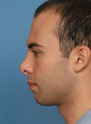
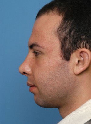
Procedure Detail: This 32 year old male presented for revision rhinoplasty. Following his original surgery by another surgeon, the patient was concerned with the appearance of the nose. Specifically, he wished to address the upturned nasal tip (over-rotation), fullness of the area above the nasal tip on profile (supratip fullness; also known as a “pollybeak deformity”), a scooped-out appearance of the profile (saddle nose deformity), and excessive nostril show. As well, the patient reported nasal obstruction. During consultation, Dr. Batniji recommended a revision rhinoplasty and performed the following:
- Revision rhinoplasty via an external (open) approach.
- Bilateral spreader grafts to address bilateral internal nasal valve collapse. During the initial rhinoplasty, a dorsal hump was removed and the nasal bones were then mobilized with osteotomies to provide refinement of the bony pyramid. However, one potential result of such a maneuver is narrowing of the internal nasal valve, resulting in excessive narrowing of the middle part of the nose (inverted “V” deformity) and nasal obstruction. Therefore, Dr. Batniji usually reinforces the middle part of the nose with spreader grafts to prevent this from happening at the time of primary rhinoplasty surgeries that he performs. In this patient’s case, he had primary rhinoplasty by another surgeon and the middle part of the nose was not reinforced. As such, Dr. Batniji performed bilateral spreader grafts in this case to improve both the appearance and function of the nose.
- Dorsal augmentation to address the scooped-out appearance of the profile. Dr. Batniji uses cartilage from the patient to perform this augmentation. While he prefers to use cartilage obtained from the septum for this augmentation, in revision cases such as this one, previous septoplasty may result in a lack of septal cartilage available for grafting. Therefore, Dr. Batniji may use cartilage from the patient’s ear to augment the dorsum and/or tip of the nose as well as use that cartilage for grafting specific areas of the nose. Dr. Batniji obtains the ear cartilage from a small incision behind the ear; removal of ear cartilage does not change the appearance of the ear. Ear cartilage was used to graft the nasal dorsum in this case.
- Sculpting of the supratip region to address the supratip fullness (“pollybeak deformity”).
- Nasal tip refinement with suture techniques and augmentation with cartilage grafting; these maneuvers enabled Dr. Batniji to correct the upturned appearance of the nose and correct the excessive nostril appearance.


